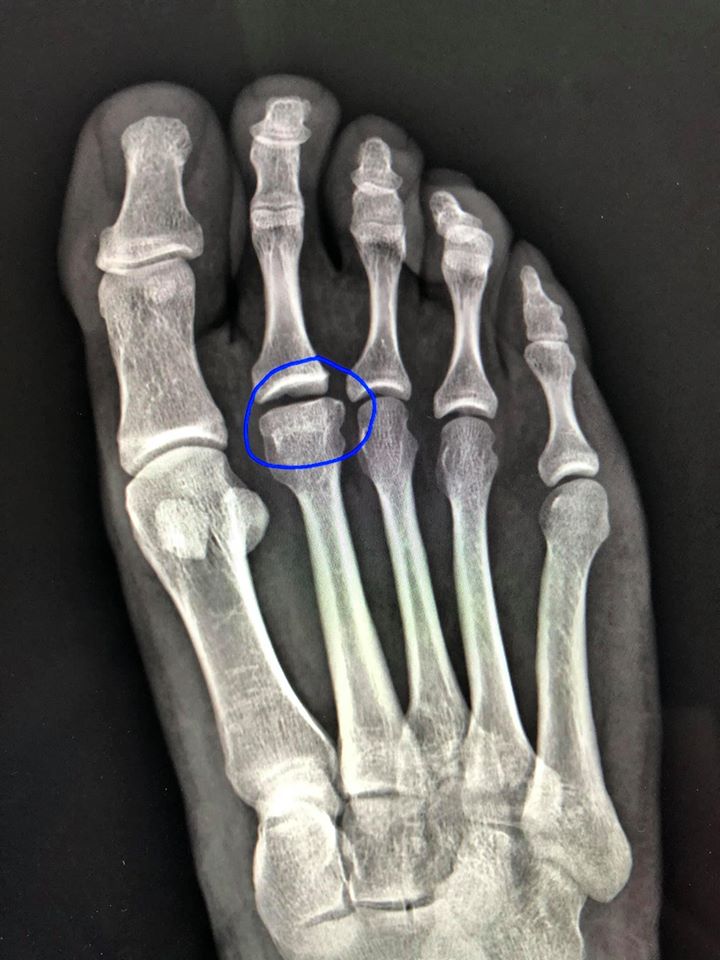
Freiberg’s disease occurs when the rounded end or ‘head’ of a metatarsal bone collapses and flattens. It most commonly affects the second and sometimes third metatarsal in the ball of the foot. It usually begins in adolescence in females who participate in activities that place repetitive stress on the forefoot such as dancing, running and athletics.
Symptoms
Freiberg’s disease is a type of osteonecrosis that affects the bone in the ball of the foot, primarily the second metatarsal. It most commonly affects young girls and adolescents during the adolescent growth period, although people of any age can develop it. Symptoms include pain, swelling, and stiffness of the foot. These symptoms are typically triggered by weight-bearing activities, particularly walking.
The exact cause of this condition is not known, but it may be the result of trauma, altered foot biomechanics, or systemic disorders that can disrupt blood supply to the affected area. It is also more apt to occur in individuals with a short second metatarsal and/or high-arched feet.
X-rays and MRI scans can help to identify this condition. Upon diagnosis, podiatrists can offer non-operative treatment to decrease the symptoms and delay progression of the disease. This includes padded inserts and custom shoes that take pressure off of the affected area when walking. Corticosteroid injections may be used to reduce inflammation and pain during flare-ups. Surgical intervention may be needed for patients with advanced Freiberg’s disease if conservative treatments are ineffective.
Surgical options include open debridement, osteotomy, and osteochondral grafting. Smith et al. 25 demonstrated positive clinical outcomes with an extra-articular metatarsal shortening osteotomy for Stage IV Freiberg’s disease. This procedure removes the avascular portion of the affected metatarsal head, which is then stabilized with a dorsal plate.
Diagnosis
This course explores the nuances of osteonecrosis (death of bone tissue) affecting the metatarsal head, most commonly the second metatarsal. It is commonly seen in adolescent girls who are participating in activities that put repetitive stress on the forefoot and may progress to pain, collapse and degeneration of the affected metatarsal head.
The cause of Freiberg infraction is unknown; however, it is believed that the condition is initiated by repetitive stress that disrupts blood supply to the growth plate of the end of the second metatarsal bone. This causes small injuries to the growth plate that eventually lead to vascular compromise and loss of bone tissue. It is also thought that footwear, such as high heels or ill-fitting shoes, contribute to the development of Freiberg infraction. Other contributing factors include anatomical variations, biomechanical stresses, and systemic disorders that influence bone metabolism or vascular health.
Symptoms of Freiberg infraction include pain that occurs with weight bearing activity, swelling in the forefoot, and stiffness or reduced range of motion in the forefoot. Diagnosis includes physical examination and x-rays of the foot. MRI or CT scans may also be useful in evaluating soft tissue and bone changes. Nonoperative treatment involves reducing foot pressure and unloading the metatarsal head with the use of orthotics, rest, crutches and activity modification. Surgical options include dorsal closing wedge osteotomies, osteochondral transplantation and resection arthroplasty.
Treatment
Freiberg disease is a rare yet clinically significant condition characterized by osteonecrosis of the metatarsal heads, most often in the second metatarsal. This osteonecrosis causes the flattening of the metatarsal head and eventual collapse, precipitating degenerative changes in the first metatarsophalangeal (MTP) joint and arthritic manifestations.
Although Freiberg disease occurs at any age, it is most common in young female athletes who engage in activities that place repetitive stress on the forefoot. This type of trauma results in a disruption of the blood supply to the end of the metatarsal bone, causing avascular necrosis. This subsequently leads to sclerosis, flattening, and collapse of the metatarsal head.
There are several theories as to the cause of this condition. Some believe that repetitive microtrauma to the metatarsal head, especially the second one, may lead to vascular compromise. Other experts suggest that anatomical variations such as a relatively long second metatarsal can lead to increased mechanical stress and predispose to the development of Freiberg disease.
Conservative treatments include pain management, activity modification, and plantar foot orthotics. If these fail to relieve symptoms, surgical treatment options are available. The gold standard surgical treatment is the Gauthier osteotomy technique, which restores sphericity of the metatarsal head and joint space, resulting in long-term clinical improvement. This procedure is especially effective in Smillie stage III and IV Freiberg disease, as it re-establishes the epiphysis covered by healthy cartilage and has no nonunion complications.
Prognosis
Freiberg disease is a rare condition characterized by osteonecrosis (death of bone tissue) of the metatarsal head, most often in the second metatarsal bone. It typically occurs in adolescents and young adults, particularly in females who participate in sports or activities that put repetitive stress on the forefoot.
Symptoms begin gradually and include pain under the ball of the foot, usually on one side and worse with activities that bend the toe. X-rays and other diagnostic tests can help your doctor determine if Freiberg infraction is present.
The exact cause is not known, but it is thought that repetitive stress causes micro-fractures to the end of the second metatarsal bone near its growth area. This can block blood flow to the bone and lead to death of tissue. The bone then changes shape and becomes more square at the end – like a “square peg” trying to fit into a “round socket.”
Treatment options depend on the stage of Freiberg disease. If diagnosed and treated early, many patients experience good results with non-surgical treatment including activity modification and a physical therapy program. For advanced Freiberg disease, surgical intervention may be necessary to improve pain and function. Surgery options include a shortening osteotomy or dorsiflexion osteotomy. Kim et al. reported that 15 feet with late-stage Freiberg disease underwent DFO and 13 of those feet had good results.