The accessory navicular is an extra bone that develops on the inside of the foot near the arch. While many people have this condition and never experience pain, in some cases the growth can irritate the posterior tibial tendon resulting in symptoms.
These symptoms can include pain with walking, swelling and redness of the middle arch of the foot. Treatment options are successful and patients can resume normal activities once they recover.
Symptoms
The accessory navicular is an extra bone located on the inner side of the foot near the arch (mid-foot area). The condition may go unnoticed and untreated until it causes pain or discomfort. Symptoms include a bump or elevated area on the inside of the foot; pain, tenderness, or swelling near the arch; and difficulty walking or engaging in physical activities due to the pain and inflammation.
It is unclear what causes this condition, but experts believe that it occurs when the bone and connective tissue develop in the foot in an abnormal manner. The condition can be diagnosed by taking a history of symptoms and performing a complete physical examination. X-rays, CT scans, and MRI may be required to identify the presence of the accessory navicular bone.
Treatment of a painful accessory navicular typically begins with nonsurgical measures. These may include a modification of activities, use of supportive footwear with orthotic inserts, and nonsteroidal anti-inflammatory medications to reduce pain and swelling. If these treatments are not effective, a temporary immobilization of the foot with the use of a cast or medical boot may be recommended to allow the area to heal. Surgery is considered if nonsurgical treatment fails to control your symptoms and interferes with your daily activities or participation in sports. Surgical removal of the navicular bone can provide relief from pain and reduce the likelihood of future problems.
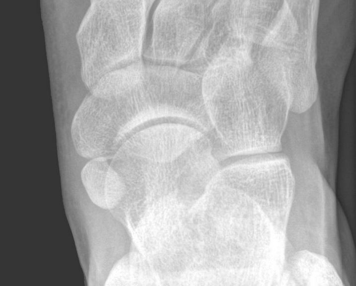
Diagnosis
A doctor can diagnose accessory navicular syndrome through a physical exam of the foot and ankle. He or she will ask the patient about any symptoms and may gently press on the area to assess for pain and tenderness. An X-ray can confirm the presence of the extra bone, and additional imaging studies like an MRI or CT scan may be required if inflammation is suspected.
Symptoms of accessory navicular syndrome typically appear after an injury or overuse and can affect one or both feet. Most often, a swollen bump appears on the inner side of the foot or arch and is irritated by shoes or aggravated by activity. This irritation is due to an over-exertion of the posterior tibial tendon, which normally supports the arch of the foot.
While the exact cause of accessory navicular syndrome is unknown, theories include incomplete joining or abnormal separation of bones and connective tissue during development. It is common for symptoms to develop during adolescence, as many teenagers begin to exercise and become more active. However, symptoms can also start to appear at any age and in adults of any gender.
Those with painful accessory navicular syndrome can usually find relief with nonsurgical treatment options. These include wearing comfortable, supportive shoes, icing the affected foot and taking nonsteroidal anti-inflammatory medications such as ibuprofen to help reduce inflammation and swelling. If the symptoms do not improve, surgery to remove the accessory navicular can provide pain relief and allow patients to return to their usual activities.
Treatment
A common foot condition that often does not cause symptoms, the accessory navicular occurs when one of the small bones in the upper inside of the foot (the navicular) develops an extra bone or ossicle. The navicular is part of the tarsal bones, which along with the calcaneus, talus, cuboid and three cuneiforms form a strong weight-bearing platform that is necessary for walking. An accessory navicular is present from birth and can affect the function of the foot and lead to pain if it becomes irritated or inflamed.
The most common symptom of an irritated or inflamed accessory navicular is pain and tenderness on the inner arch of the foot, which can be exacerbated by activities that put stress on the arch. The area can become red and swollen, and may feel sensitive to touch. Ill-fitting footwear can also aggravate the discomfort.
Non-surgical treatment is usually the first option, and aims to reduce inflammation and pain. This can include resting the foot and reducing high-impact activities, the use of ice and nonsteroidal anti-inflammatory medications such as ibuprofen, and custom orthotic inserts that provide support to the arch of the foot. If non-surgical treatment fails to improve the symptoms, surgical removal of the accessory navicular is recommended. This is a minimally invasive procedure performed under local anesthesia and involves detaching the accessory navicular from the posterior tibial tendon, which allows the bone to heal and decreases pressure on the navicular and arch of the foot.
Prevention
The navicular bone is a boat-shaped bone located in the arch of the foot. Often, an extra bone or cartilage develops near this bone during development, forming a condition known as accessory navicular. In many cases, the presence of an accessory navicular is asymptomatic and does not cause pain. However, in some individuals, this bone can irritate the posterior tibial tendon and lead to symptoms. Several factors can contribute to the occurrence of accessory navicular syndrome, including a genetic predisposition and variations in foot structure.
Symptoms of accessory navicular syndrome are typically mild and can be managed with non-surgical treatment. This typically involves rest, activity modification, ice application, and the use of custom orthotic devices to improve foot function and reduce stress on the midfoot. Physical therapy is also frequently recommended to strengthen the foot muscles and other soft tissues in the area.
Despite its relative rarity, the presence of an accessory navicular can significantly impact one’s daily functioning. Therefore, it is important to seek routine evaluations with a podiatrist or orthopedic specialist. This will ensure that early diagnosis and effective management of this condition are possible. By combining a thorough examination, customized treatment plans, and preventive measures, it is possible to manage the symptoms of ANS effectively. This will allow you to maintain optimal foot health and continue the activities that are important to you.