Calcaneal apophysitis, also called Sever’s disease, is heel pain at the back of the heel bone (calcaneus). This occurs in children between the ages of 8 and 14 years during periods of growth spurts.
During this time the bones grow faster than the muscles and tendons. This causes stress to be placed on the growth plate, which can cause inflammation.
Causes
Calcaneal apophysitis, is a painful inflammation of the heel bone (calcaneus) growth plate in children. Growth plates are areas at the ends of bones that undergo changes during childhood to allow for bone growth. This area is weaker than the surrounding bone and tendons and is vulnerable to repetitive stress such as in jumping or running. The condition is common in children during their growth spurt and occurs in both feet.
The pain is typically worse at the end of a physical activity and subsides when the activity is stopped. The pain may also be aggravated by walking on hard surfaces or wearing shoes that have a higher heel than normal (such as soccer boots). Other factors that can lead to calcaneal apophysitis include changing from a low to high impact sport, being overweight, walking or playing barefoot at the beach in thongs/flip flops.
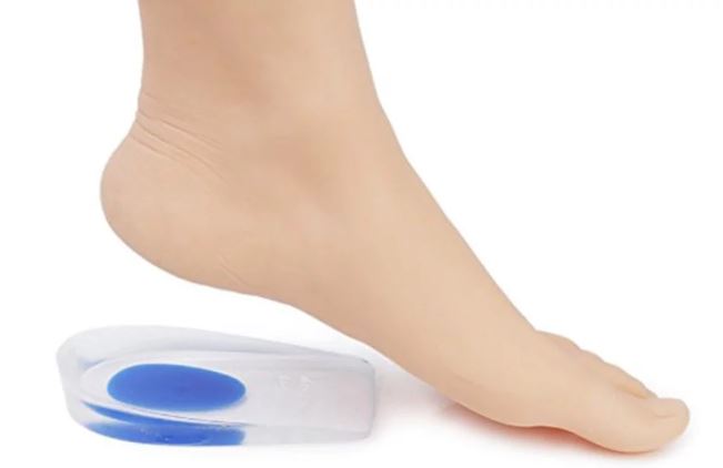
Most children with calcaneal apophysitis do not improve with rest and are unable to walk, run or play without pain. This may lead to poor fitness and social activities and emotional distress in some children. It is important that this condition is diagnosed early and treated effectively. Treatment options include icing, stretching and shock-absorbing heel inserts. In a randomized controlled trial, shock-absorbing heel inserts were found to be effective in reducing the symptoms of calcaneal apophysitis in children between eight and fifteen years of age.
Symptoms
Calcaneal apophysitis is painful irritation and inflammation of the growth plate located at the back of the heel bone. The growth plate, which is made of cartilage, is weaker than the solid bone of the heel and is prone to injury from repetitive stress. The condition typically affects children between ages 8 and 14, especially those who participate in sports that involve running and jumping.
Children who are experiencing calcaneal apophysitis tend to be taller and heavier, thus placing more stress on the growth plate of the heel. They often play sports that require repetitive running and jumping, such as soccer and basketball, or have recently started a new sport.
The pain from calcaneal apophysitis is most severe after prolonged activity and when pressure is applied to the heel. It also becomes more intense when your child is standing on their feet all day, such as when at school or work. They may limp or walk on their tiptoes to relieve the pressure on the heel. The pain improves with rest and usually returns with activity. A physical examination by a podiatrist and imaging tests, such as X-rays, are usually sufficient to diagnose the condition. In severe cases, a cast may be used to promote healing and keep the foot immobile. This can help reduce the risk of recurrence of the condition.
Diagnosis
The diagnosis is primarily made by history and physical exam. Affected children often present with pain during or after sports activities and may report it has been getting worse. Children who participate in sports that require repetitive running and jumping are at a higher risk of developing this condition.
Heel pain in adolescents and children is common and most often caused by mechanical overuse. However, it is important to distinguish calcaneal apophysitis from other conditions that share clinical features and are commonly confused with Sever disease, including Achilles tendon rupture, calcaneal spurs, Achilles tendinopathy, retrocalcaneal bursitis, fifth metatarsal apophysitis, and osteomyelitis.
An active 12-year-old boy presents to your office with heel pain that occurs most often during or after athletic activity. He reports it has gotten worse over time and complains that standing tiptoe makes the pain worse. You perform a passive dorsiflexion test and find that the patient has limited ankle movement. You also perform the squeeze test, a maneuver that requires mediolateral compression of the calcaneal epiphysis. This produces pain, usually in the back of the heel.
X-rays are typically normal, but can help to rule out a fracture. MRI has been reported to be helpful in identifying the area of inflammation by showing signal changes on the unossified apophysis. Historically, clinicians have restricted patients’ activities in an attempt to heal this painful condition. This has been shown to be less effective than allowing the body to heal naturally, and is considered controversial.
Treatment
The condition, also known as Sever’s disease, is pain at the back of the heel bone growth plate (calcaneal apophysis). It typically affects children between the ages of 8 and 15, especially those who participate in sports that require repetitive running or jumping.
The pain is caused by excessive pressure on the growth plate from repeated use of the calf muscles and the Achilles tendon. This stress irritates the unossified apophysis, leading to microtrauma and thickening of the area. This pain is most common in athletes who play sports like soccer, football and basketball that involve frequent running or pivoting movements. It may also occur in those who experience rapid growth spurts or have a foot biomechanical problem such as flatfoot, a tight Achilles tendon or high-arched feet.
Although pain in the heel is a very common injury that requires treatment, it’s important to note that there are many other conditions that mimic calcaneal apophysitis and need to be differentiated from it. Therefore, it is important to have your child thoroughly evaluated by a doctor who has extensive experience with children’s injuries.
A physical exam typically includes palpation of the heel and foot to locate the area of the tenderness. In addition, a passive dorsiflexion test, which involves gently pushing on the bottom of the foot while your child is standing tiptoe, and a squeeze test of the lower one-third of the posterior calcaneal epiphysis can help confirm the diagnosis of calcaneal apophysitis.